Nomogram for Giant Cell Tumor of Bone
骨巨細胞腫のノモグラム

RESEARCH
研究紹介
![]()
RESEARCH
研究紹介
Orthopedic surgeons perform curettage (joint-preserving surgery) rather than en bloc resection for bone giant cell tumors of the extremities in order to achieve good functional outcomes; however, a high local recurrence risk remains a concern. If the risk of local recurrence is adequately understood, careful examination and frequent imaging studies can be performed in high-risk patients, enabling early detection of local recurrence and subsequent curettage or radiofrequency ablation, thereby preserving the joint. We have developed a web-based nomogram to predict the risk of local recurrence after curettage of bone giant cell tumors of the extremities. This nomogram can be provided to clinicians to enhance their ability to assess patient prognosis, strengthen prognosis-based decision-making, improve patient stratification, and inform patients in the clinic. External validation using data from other institutions is required in the future.
This nomogram has been developed by Masunaga T, Tsukamoto S, Kurakami H, Honoki K, Fujii H, and Kido A from Nara Medical University, Nara, Japan and Donati DM and Errani C from IRCCS Istituto Ortopedico Rizzoli, Bologna, Italy.
The nomogram is intended solely to provide reference information based on statistics. When making actual treatment decisions, we recommend consulting with your attending physician. Our institutes shall not be liable for any disadvantages arising from the use of this nomogram. Use of this nomogram and the results of calculations are at your own risk. We encourage the user to rely on the published papers for details.
| Age | |
|---|---|
| Site | |
| Campanacci stage | |
| Pathological fracture at presentation | |
| Preoperative denosumab therapy | |
| Previous surgery | |
| Filler | |
| Local adjuvant therapy |
0%
0%
A nomogram for predicting the local recurrence risk after curettage for giant cell tumor of bone in the extremities before treatment begins is available here.
| Age | |
|---|---|
| Site | |
| Campanacci stage | |
| Pathological fracture at presentation | |
| Previous surgery |
0%
0%
(Cited from Mavrogenis AF, Igoumenou VG, Megaloikonomos PD, Panagopoulos GN, Papagelopoulos PJ & Soucacos PN (2017) Giant cell tumor of bone revisited. SICOT J, 3, 54)
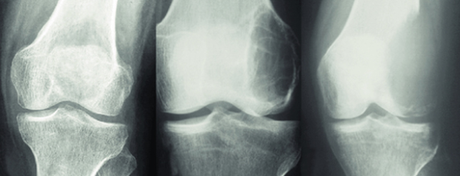
Campanacci stage is most often used for stage classification of giant cell tumor of bone and is defined as follows. Stage I tumor has a well marginated border of a thin rim of mature bone and the cortex is intact or slightly thinned but not deformed. Stage II tumor has relatively well-defined margins but no radio paque rim; the combined cortex and rim of reactive hone is rather thin and moderately expanded but still present. Stage III tumor designats a tumor with fuzzy borders, suggesting a rapid and possibly permeative growth; the tumor bulges into the soft tissues. But the soft-tissue mass does not follow the contour of the bone and is not limited by an apparent shell of reactive bone (Campanacci M, Baldini N, Boriani S, Sudanese A. Giant-cell tumor of bone. J Bone Joint Surg Am. 1987;69:106–14.).
Local adjuvant therapy refers to the treatment of the cavity after curettage of the giant cell tumor of bone, followed by phenol and alcohol, cauterization with argon beam coagulator, or freezing with liquid nitrogen spray.